Microscopy New Zealand
Welcome to Microscopy New Zealand's Website
We hope you enjoy our contribution to the pursuit of microscopy
Welcome to Microscopy New Zealand's Website
We hope you enjoy our contribution to the pursuit of microscopy
Lincoln University 2nd-6th December 2024.
See more details here.
Anyone with an interest in microscopy is welcome to join Microscopy New Zealand, the only entry criterion is enthusiasm and an occasional willingness to participate in the society.
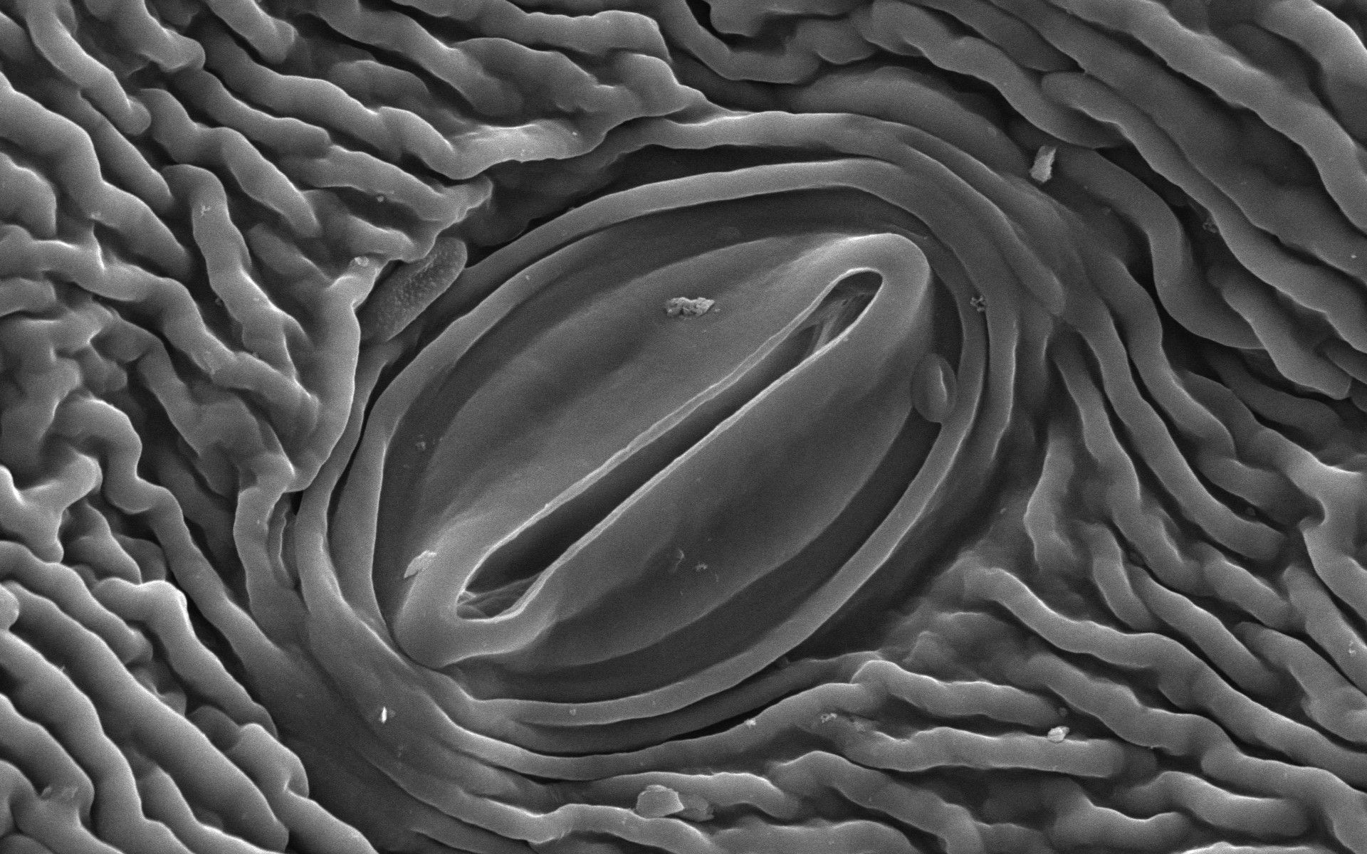
Scanning Electron Microscope image of stomatum
Most of the members work at universities scattered across New Zealand, others at Crown Research Institutes or private industry. Microscopy is a tool that is useful in many situations, so it's fair to say there is a wide range of uses our society members put them to.
The equipment we use ranges from simple desktop stereo microscopes, through to multi-photon confocal microscopes; scanning and transmission electron microscopes; and even a few Micro-CTs for good measure.
The intention of MNZ is to keep the people using this equipment in touch with with each other and help develop collaboration and general enthusiasm for the equipment and techniques we use.
New Zealand has a low population and apart from Auckland, most of it is scattered thinly across the country. As New Zealand doesn't possess an unusual national passion for microscopy, it stands to reason that our microscopy community is also very small. So we're always on the lookout for international members to join our society and exchange their experiences with us.

From Microscopy Australia's website: "It our great pleasure to announce a new microscopy exchange scheme between Australia and New Zealand established in partnership with the Australian Microscopy and Microanalysis Society (AMMS) and Microscopy New Zealand (Microscopy NZ)."
Opportunities to be hosted at other facilities, or host staff from them in your own are often the best ways to broaden knowledge and form long-term collaborations. Sure, social media, Zoom and email are great in their own ways, but working with real people beats them all. To learn more the details, head to Microscopy Australia's link: https://micro.org.au/news/microscopy-knowledge-exchange-visits/
Please Note: Due to the ever-changing Covid environment, there may be changes to the proposals, or when exchanges can take place. Every effort will be made to accommodate any such obstacles.
If you have any questions not answered here, please contact Associate Professor Mihnea Bostina, University of Otago.
Page Banner Credit
BPAE cells, 20x, Labelled with MitoTracker (green), phalloidin (red), and DAPI (blue), confocal. Theano Stafidas, ATA Scientific Australia.